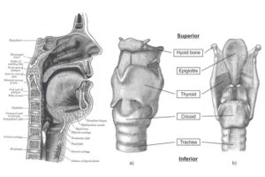
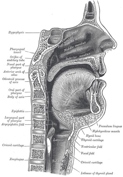
The vocal cords, also known as the vocal folds are two band-like structures that are located within the larynx (voice box) above the trachea and responsible for voice production. They are widely referred to as vocal folds because they seem more like folds of tissues than cords. The vocal cords are located midway in the trachea or throat and are protected by the thyroid cartilage also known as Adam’s apple.
How do the vocal cords work?
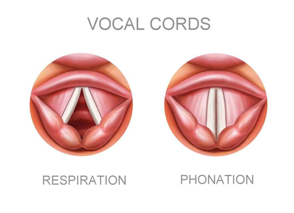
The vocal cords stay open during inhalation and close up during phonation and swallowing. The production of sound is as a result of the vibration of the chords as they respond to the passage of exhaled air between them. When the vocal cords are closed, they may vibrate and modulate the airflow expelled by the lungs to produce vocal sounds.
The vocal cords are so small in size that they measure only between 1.75 cm and 2.5 cm (approx 0.75″ to 1.0″) in length for males, and between 1.25 cm and 1.75 cm (approx 0.5″ to 0.75″) in length for females and just a few millimeters in thickness. The vocal cords are thinner and shorter in children and women; this explains the higher pitched voices of women and children. The size and color of the vocal folds differ in males and females. While adult males typically have longer and larger cords that lead to lower-pitched voices, the vocal cords found in women are thinner, shorter and more pearly white.
Anatomy of the vocal cords
The anatomy of the vocal cords are quite complex and it is made up of three layers. The layers include:
- The epithelium which is the outer “skin” layer
- The lamina propria, it has three sub layers, which are;
Deep; this is the collagenous layer
Intermediate; this is the elastic layer
Superficial; this layer has the consistency like that of a jelly
- The vocalis muscle also known as the thyroarytenoid muscle.
The superficial layer of the lamina propria and the epithelium are what make up the mucosa layer which is gel-like and soft. The vocal ligament is made up of the deep and intermediate layer of the lamina propria while the vocalis muscle is the stiff and major body of the vocal fold.
GREAT BOOKS WE RECOMMEND, CLICK ON THE IMAGE TO KNOW MORE!
Anatomy of how the vocal cords function
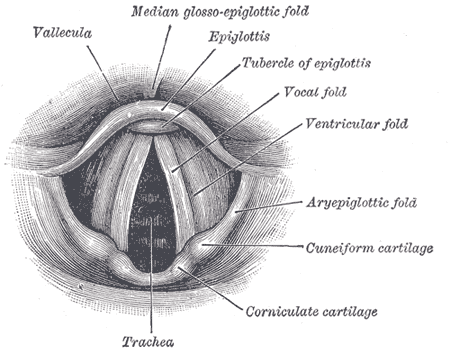
The vocal cords project posteriorly to the arytenoid cartilage found on both sides and are attached anteriorly at an angle to the interior surface of the thyroid cartilage. The superior layer of infolded membrane are responsible for the formation of the ventricular folds also known as the false vocal cords ( or vestibular folds ) while the true vocal cords are formed from the inferior layer of the infolded membrane. The most important function of the false vocal cords is the protection of the very delicate vocal cords lying just beneath it. The false vocal cords play a minimal role on phonation but could be used in throat singing, screaming or the production of deep tones.
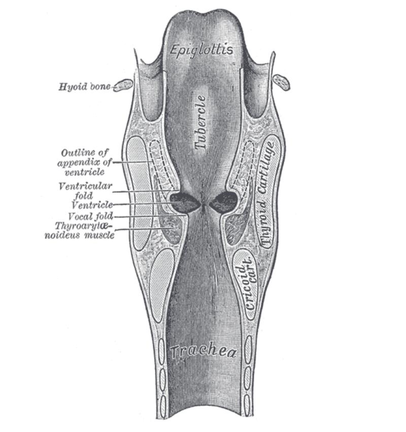
On both sides of the middle laryngeal cavity and in between the vocal cords and the false vocal cords, there is a machine that bulges laterally to form troughs that are known as laryngeal ventricles. The laryngeal saccules are located between the vestibular false vocal cords and the thyroid gland, and they’re tubular extensions of each ventricles. Importantly, the walls of the saccules are believed to have a lot of mucous glands that help to lubricate the vocal cords.
There’s a free superior margin of the submucosal membrane that is thickened to form the vocal ligament that forms the true vocal cords as soon as it’s covered by mucosa. The function of the muscle fibres found in the true vocal folds is for fine tonal control of the vocal cords.